Sutter’s CPMC Performs Its First AI-Guided Vascular Fusion Procedure
By Jenn Lonzer, Vitals contributor
Every morning before the sun climbed high in the sky, Maria De La Luz Velasco walked.
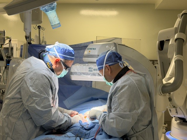
Dr. Hernan Bazan (left) and Dr. James Wong (right) treat Maria De La Luz Velasco
An 86-year-old grandmother, De La Luz Velasco always started her day circling the football field near her home. She preferred the cool, quiet hours before the daylight grew too bright — strong sunlight hurt her eyes.
Then came the abdominal pain.
For a little over a week, De La Luz Velasco had felt sharp abdominal pain, especially after eating. When it became unbearable, she went to her local emergency department. There, a CT angiogram revealed the problem: the arteries supplying blood to her intestines were severely blocked. Without treatment, this condition — known as acute-on-chronic mesenteric ischemia — can progress to bowel infarction, a life-threatening emergency.
De La Luz Velasco was transferred to Sutter’s CPMC in San Francisco, where she met with Dr. Hernan Bazan, chief of vascular & endovascular surgery. With a history of coronary artery bypass surgery and heart failure, De La Luz Velasco was an exceptionally high-risk surgical candidate. A traditional open operation would have placed tremendous stress on her heart and body.
Instead, Dr. Bazan recommended a minimally invasive endovascular procedure. Working through a small puncture in De La Luz Velasco’s groin, he would navigate wires, catheters and stents through her blood vessels to restore blood flow to her intestines.
But before De La Luz Velasco ever entered the operating room, the procedure had already begun.
Building the Roadmap
In the control room overlooking the hybrid operating room, Dr. Bazan and his team, including his partner Dr. James Wong, gathered around a bank of computer monitors.
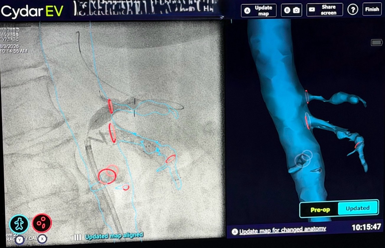
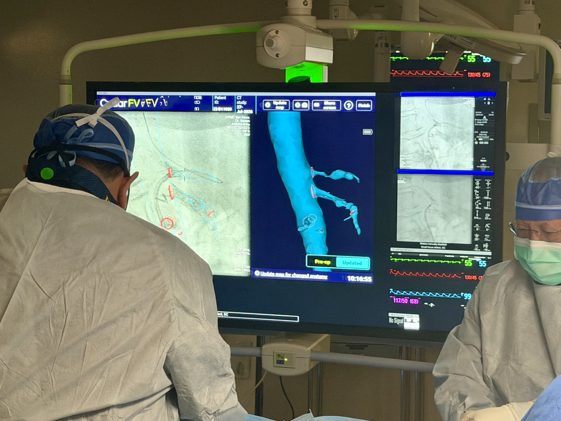
Red targets mark the entrances to De La Luz Velasco’s arteries
On the screens, De La Luz Velasco’s anatomy slowly rotated in three dimensions.
Her spine.
Her aorta.
The arteries branching toward her intestines.
The model had been created from her CT scan using a “Cydar map,” an AI-guided fusion software that transforms conventional imaging into a patient-specific, three-dimensional reconstruction.
Dr. Bazan turned the model again and again, studying it from different angles.
Then the surgical team began placing small red targets.
They weren’t marking the disease.
They were marking the path to it.
Each target identified the tiny opening where an artery branched from the aorta. Before a blockage could be treated, Dr. Bazan first had to find and enter that opening — one of the most technically demanding steps in the procedure.
The team discussed the best projection angles for imaging, which catheters would be most likely to engage the heavily calcified vessels and how they would approach each blockage.
Only after every step had been carefully planned did De La Luz Velasco enter the operating room.
Finding the Way
Inside the hybrid OR, the atmosphere changed. The lights dimmed and large monitors flickered to life.
After accessing the artery in De La Luz Velasco’s groin, Dr. Bazan advanced a guidewire toward her aorta.
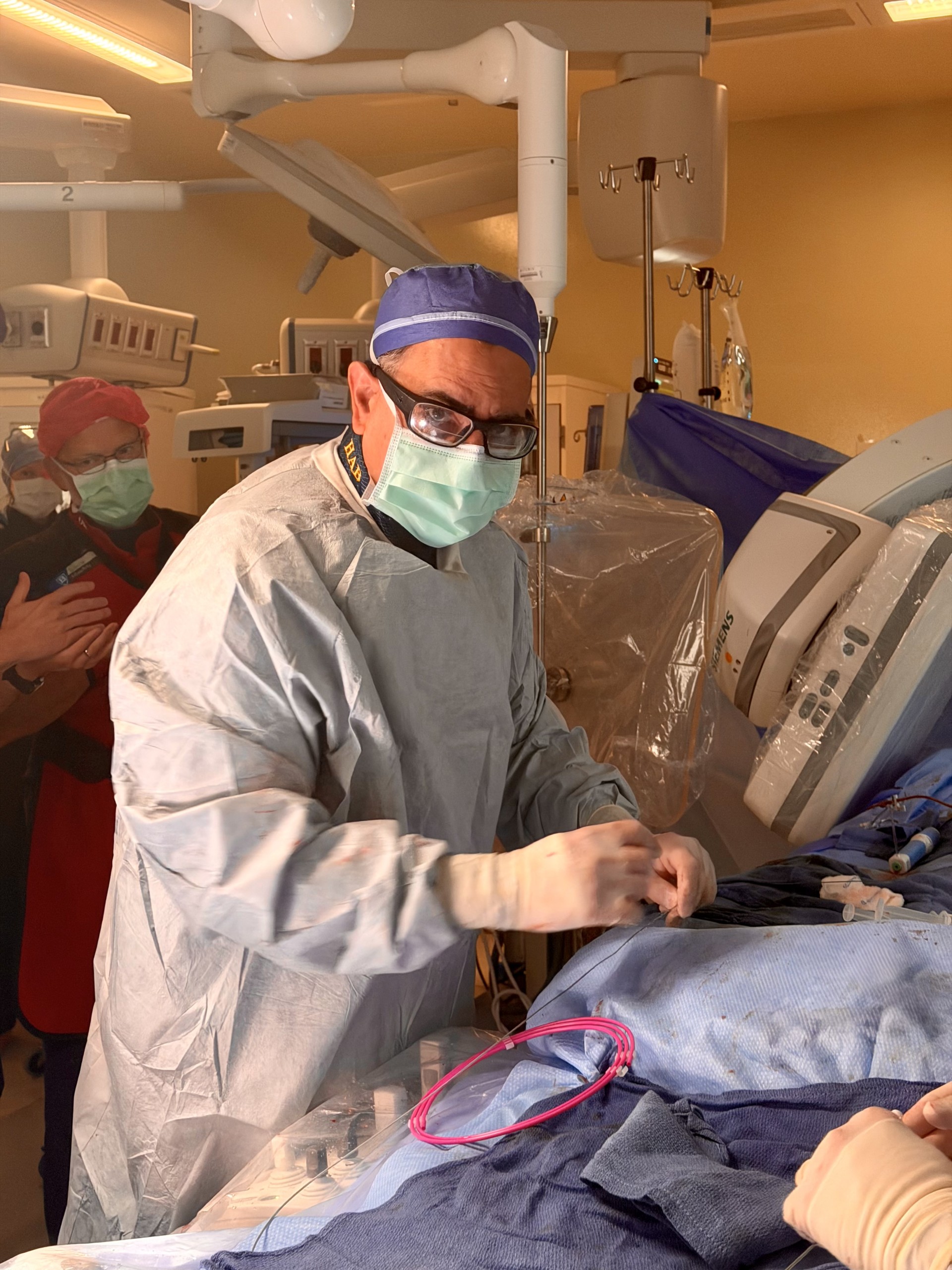
Dr. Hernan Bazan performs a minimally invasive endovascular procedure on Ms. De La Luz Velasco at Sutter’s CPMC in San Francisco
The anatomic model from the planning room appeared over live fluoroscopy — the real-time X-ray surgeons use to guide wires and catheters through blood vessels. The same red targets marking the entrances to De La Luz Velasco’s arteries were waiting for him.
Before fusion imaging, surgeons relied on conventional angiography — requiring higher radiation exposure and contrast dye — to translate CT scans into the anatomy encountered under fluoroscopy. Some even drew targets directly on the monitor with a marker.
“It’s like driving with a heads-up GPS navigation with traffic and weather data projected right onto your windshield instead of glancing at a paper map in your lap,” Dr. Bazan said. “You’re still driving. You’re still making every decision. But the information is right where you need it, exactly when you need it.”
Unlike roads, anatomy changes during a procedure. As guidewires and catheters move through the arteries, vessels can straighten, stretch and shift slightly. A mark drawn on a monitor stays fixed. The digital targets do not. Registered to the patient’s anatomy, they move with it, helping the team stay oriented throughout the procedure.
The team still paused at key moments to confirm the position of guidewires, catheters and stents. Each time, the ceiling-mounted imaging system swept over the table. Dr. Bazan positioned a clear lead shield near De La Luz Velasco’s chest and asked the anesthesiologist to briefly hold her breathing while new X-ray images were captured.
Those images remained an essential safety check. What changed was their purpose. Instead of relying on repeated X-rays to find the openings of De La Luz Velasco’s arteries, Dr. Bazan used the AI-guided model to navigate to them. Fluoroscopy then confirmed that each device was exactly where it needed to be before the procedure moved forward.
AI helped the team navigate. Fluoroscopy confirmed they had arrived.
A Procedure Measured in Millimeters
Dr. Bazan and his team first crossed the superior mesenteric artery — the primary vessel supplying the intestine — and placed a stent to open it, restoring strong blood flow. The second target, the celiac artery, which feeds the upper digestive organs, presented an even greater challenge due to severe calcium buildup.
As the team worked it into position, the stent partially slipped from the balloon designed to carry it.
The room didn’t become frantic.
It became quieter.
Working methodically, the team reassessed, repositioned the devices, and carefully recovered the stent without damaging the artery. After deploying a second stent to fully treat the blockage, blood flow was restored.
By the end of the procedure, both major arteries supplying De La Luz Velasco’s intestines were open once again.
The two arteries presented very different challenges, revealing where AI-assisted fusion imaging fits into today’s operating room.
During preoperative planning, Dr. Bazan used the technology to identify the tiny openings where the arteries branched from the aorta. In the operating room, those digital targets helped guide the team to the vessel origins with greater confidence. But once inside the arteries, the challenge shifted. Crossing heavily calcified blockages, selecting the right devices and responding when the unexpected happened still depended entirely on surgical judgment, experience and teamwork. “This technology gives us an incredible map to get to the front door,” Dr. Bazan noted. “But once you’re inside a heavily calcified vessel, judgment, team communication, and decades of experience take over.”
An AI First for Sutter Health
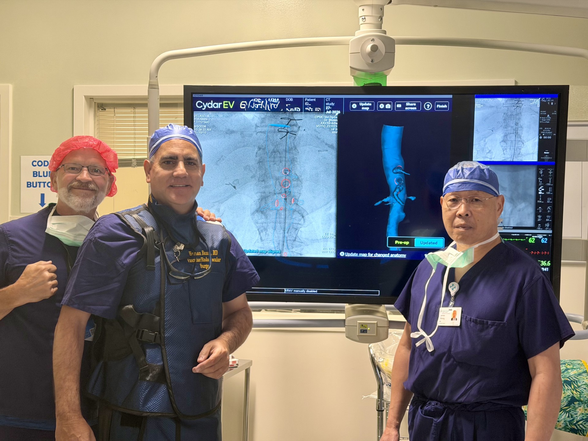
Dr. Greg McIff (Cydar Medical), Dr. Hernan Bazan (center) and Dr. James Wong (right)
De La Luz Velasco’s case represented more than a successful operation. It marked the first use of AI-assisted fusion imaging at Sutter.
“This is a significant step forward for our surgical capabilities,” says Dr. Bazan of Sutter West Bay Medical Group. “My hope is to make it scalable and ultimately the standard of care across Sutter’s tertiary hospitals.”
Because the cloud-based platform has already completed Sutter’s IT, cybersecurity and AI governance review, Dr. Bazan believes the groundwork has been laid to expand its use across the organization.
At Sutter’s CPMC, the team also plans to use the technology to support the surveillance and treatment of abdominal aortic aneurysms, where patient-specific 3D planning can help guide increasingly complex endovascular procedures.
The Measure of Success
The day before surgery, De La Luz Velasco wasn’t eating. Following the procedure, she was eating solid food again, and follow-up CT imaging confirmed that both mesenteric stents remained wide open and positioned perfectly.
The operation required meticulous planning, advanced imaging, evolving technology, and countless decisions made by an experienced surgical team. Yet its ultimate success wasn’t measured by the AI model, the fluoroscopy images, or the stents.
It was measured at breakfast — and in an 86-year-old grandmother recovering well and heading home to her family.